Revolutionary MRI Imaging Technique Reveals Brain Glucose Metabolism Without Radiation Exposure
A new MRI imaging technique has been developed by a research team at MedUni Vienna that can map brain glucose metabolism without the use of radioactive substances. The non-invasive procedure uses a harmless glucose solution and can be used with common MRI scanners, offering reliable results for diagnosing metabolic disorders.
Metabolic disorders play a central role in many common conditions, including Alzheimer’s, depression, diabetes and cancer, which call for reliable as well as non-invasive diagnostic procedures. Until now, radioactive substances have been administered as part of the process of mapping glucose metabolism in the brain.
Now, a MedUni Vienna research team has developed a completely new magnetic resonance imaging (MRI) approach. Using a harmless glucose solution, the procedure generates reliable results and—in principle—can be used with all common MRI scanners.
The findings from the study have just been published in the journal Nature Biomedical Engineering.
The study looked at—and has significantly enhanced—current diagnostic procedures for mapping brain glucose metabolism. The results were generated by measuring blood glucose levels and metabolic products in healthy subjects several times during a period of around 90 minutes.
In contrast to existing procedures, the subjects did not receive radio-labeled glucose but a quantity of a harmless glucose solution equivalent to a can of a fizzy drink. As this substance does not produce a direct signal for the MR imaging method used, concentrations and metabolism of glucose were measured indirectly based on the drop in signal intensity for the product concerned.
“The main advantage of this indirect method is that it can be used on other MR devices without any difficulties, because no additional hardware components are required, as is the case with other, comparable approaches,” explained principal investigator Wolfgang Bogner of the Department of Biomedical Imaging and Image-guided Therapy at MedUni Vienna, highlighting the clinical significance of the research findings.
Broad range of potential applications
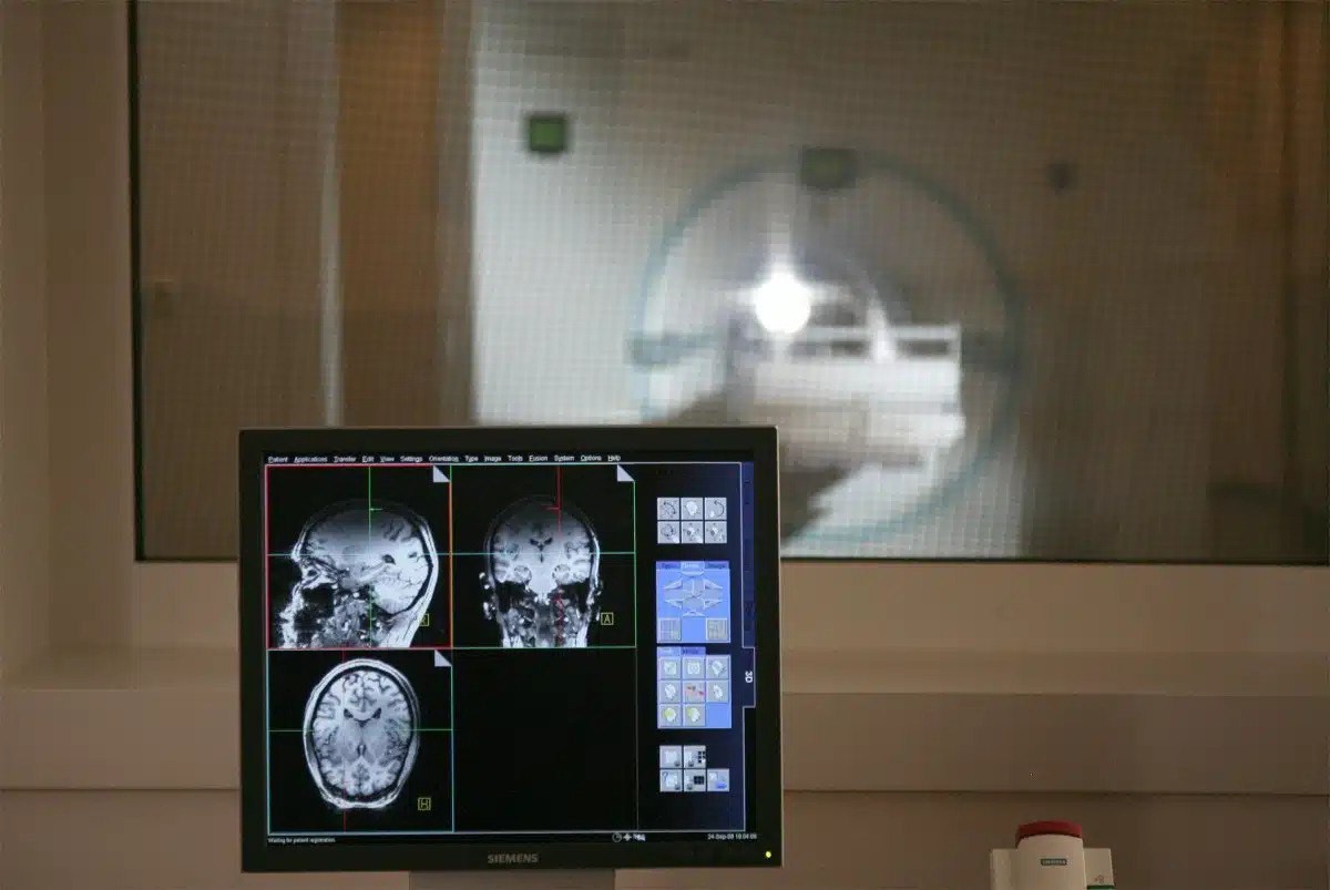
Carried out by researchers from the Department of Psychiatry and Psychotherapy and Department of Medicine III at MedUni Vienna, the study used the university’s high-performance 7-Tesla MRI scanner. The device began operation in 2008 and is still the only ultra-high-field MR scanner available in Austria. Wolfgang Bogner and his team have already demonstrated that the novel approach also works on 3-Tesla MR scanners.
Pioneering MRI imaging method captures brain glucose metabolism without the need for administration of radioactive substances. Credit: Medical University of Vienna
“That was an important step, because 3T MR systems are extremely widespread in clinical applications,” said Fabian Niess, lead author of a follow-up study published in Investigative Radiology.


Related Posts