Another analysis available to researchers in this Lab is the recording and preparation of T1 & T2 maps. The raw Relaxometry images received from the MRI device contain T1 or T2 weighted images that provide a relatively good contrast at specific TR and TE. However, with Relaxometry techniques, you can get the actual number of this tissue features and calculate the actual map (instead of the weighted image).
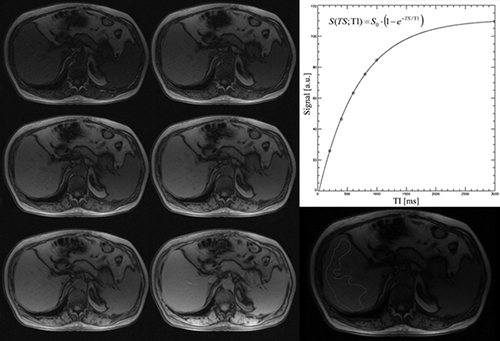
Figure 1: An example of T1 Relaxometry using the spin echo technique.
List of services:
We use MATLAB for analyzing the researcher's data. ImageJ software also provides the ability to perform this analysis using the MRI Processor plugin.