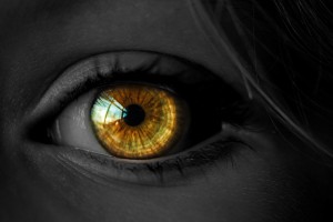
Eye could provide “window to the brain” after stroke
Research into curious bright spots in the eyes on stroke patients’ brain images could one day alter the way these individuals are assessed and treated.
Research into curious bright spots in the eyes on stroke patients’ brain images could one day alter the way these individuals are assessed and treated. A team of scientists at the NIH found that a chemical routinely given to stroke patients undergoing brain scans can leak into their eyes, highlighting those areas and potentially providing insight into their strokes.
They were kind of astounded by this – it’s a very unrecognized phenomenon. It raises the question of whether there is something they can observe in the eye that would help clinicians evaluate the severity of a stroke and guide us on how best to help patients.
The eyes glowed so brightly on images due to gadolinium during MRI scans to highlight abnormalities in the brain. In healthy individuals, gadolinium remains in the blood stream and is filtered out by the kidneys. However, when someone has experienced damage to the blood-brain barrier, gadolinium leaks into the brain, creating bright spots that mark the location of brain damage.
Previous research had shown that certain eye diseases could cause a similar disruption to the blood-ocular barrier, which does for the eye what the blood-brain barrier does for the brain. Dr. Leigh’s team discovered that a stroke can also compromise the blood-ocular barrier and that the gadolinium that leaked into a patient’s eyes could provide information about his or her stroke, so the eye is reflective of what is going on in the brain.
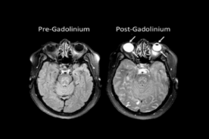
The researchers compared patient scans without and with administering gadolinium . Because gadolinium is transparent, it did not affect patients’ vision and could only be detected with MRI scans.
Gadolinium was typically present in the front part of the eye, called the aqueous chamber, after two hours, and in a region towards the back, called the vitreous chamber, after 24 hours. Patients showing gadolinium in the vitreous chamber at the later timepoint tended to be of older age, have a history of hypertension, and have more bright spots on their brain scans, called white matter hyperintensities, that are associated with brain aging and decreased cognitive function.
In a minority of patients, the two-hour scan showed gadolinium in both eye chambers. The strokes in those patients tended to affect a larger portion of the brain and cause even more damage to the blood-brain barrier than the strokes of patients with a slower pattern of gadolinium leakage or no leakage at all. The findings raise the possibility that, in the future, clinicians could administer a substance to patients that would collect in the eye just like gadolinium and quickly yield important information about their strokes without the need for an MRI.
www. nih.gov

Related Posts