Classification of Cortical Neurons by Spike Shape and the Identification of Pyramidal Neurons
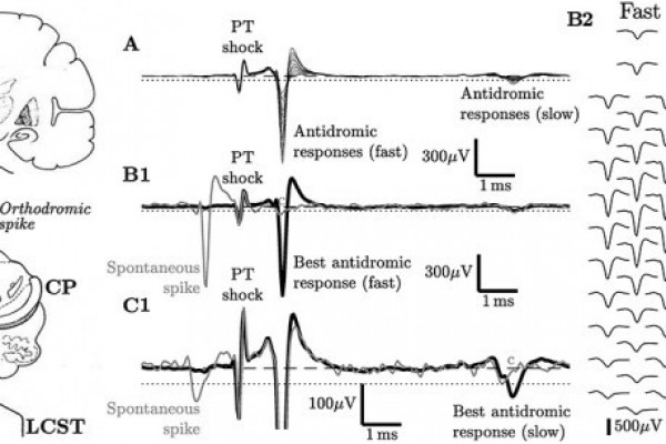
Many investigators who make extracellular recordings from populations of cortical neurons are now using spike shape parameters, and particularly spike duration, as a means of classifying different neuronal sub-types. Because of the nature of the experimental approach, particularly that involving nonhuman primates, it is very difficult to validate directly which spike characteristics belong to particular types of pyramidal neurons and interneurons, as defined by modern histological approaches. This commentary looks at the way antidromic identification of pyramidal cells projecting to different targets, and in particular, pyramidal tract neurons (PTN), can inform the utility of spike width classification. Spike duration may provide clues to a diversity of function across the pyramidal cell population, and also highlights important differences that exist across species. Our studies suggest that further electrophysiological and optogenetic approaches are needed to validate spike duration as a means of cell classification and to relate this to well established histological differences in neocortical cell types.
It is now widely recognized that understanding the operation of cortical circuits requires not only careful documentation of neuronal activity but also better identification of the neuronal cell types exhibiting that activity. Technical advances have allowed better and better insights into the activity of large populations of cortical neurons. These approaches are being actively employed in a variety of animal models, including mice, rats, and macaque monkeys, with extracellular recordings from probes carrying multiple contacts.
One of the most enduring ideas is that extracellular spike shape might help to distinguish different classes of neuron recorded in this type of study. Early investigators, recording in the awake monkey, first suggested that interneurons (stellate or Golgi type II), with high spontaneous firing rates, had “thin” action potentials of short duration and could be distinguished from putative pyramidal cells with broader spikes and a lower, regular spiking pattern of discharge. These differences were subsequently confirmed by detailed electrophysiological investigations in rodent neocortex. Other early investigations in rabbit neocortex used the antidromic response of pyramidal neurons with axons in the corpus callosum to distinguish them from other types of neuron, including interneurons.
Recent work has used a number of different spike waveform and discharge parameters to identify different classes of neuron recordings from prefrontal and posterior parietal cortex; these classes were then shown to differ in terms of their response dynamics and information coding. The classes with the briefer spikes were considered to be putative interneurons; those with the broader spikes as putative pyramidal cells.
This commentary has emphasized that where electrophysiological identification has been possible through antidromic testing, the results suggest that spike shape alone cannot provide a reliable means of differentiating at least one sub-type of pyramidal cells, the PTNs, from interneurons. We are reiterating caution on this point, first made many years ago Swadlow and repeated since Vigneswaran, our concern being strengthened by new knowledge on the range of spike widths exhibited by macaque pyramidal cells and the striking differences between rodent and macaque in the expression of at least one membrane K+ channel that helps to determine spike width. The perils of misclassification are considered by some investigators to be limited. However, given that pyramidal neurons with “thin” spikes have now been documented in more than one cortical area and in three different species (rabbit, cat, and macaque), greater caution is surely needed. In particular, the existence of PTNs with “thin” spikes in motor areas of macaque cortex should not be ignored, but rather should encourage investigation of PTNs in other cortical sensorimotor areas, and indeed of other layer V corticofugal neurons in wider regions. The use of correlation techniques for comparing the actions of pyramidal neurons with those of interneurons should also be extended. Firing rate related measures should also be included into consideration. All efforts to classify cell types using measures derived from extracellular recordings are important since it may potentially lead to the understanding of the computations performed by different classes in awake behaving animals. But we would like to emphasize that linking such classifications to anatomical, morphologically based classifications is not easy and requires caution. In the macaque, no doubt, better characterization of neuronal subtypes will be achieved using genetic dissection and optogenetic stimulation, exploiting the huge advances made in the mouse.


Related Posts