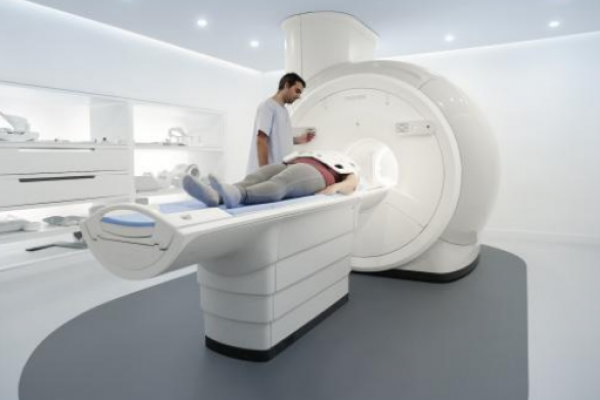
Brain Iron Levels May Predict Multiple Sclerosis Disabilities
A new, highly accurate magnetic resonance imaging (MRI) technique can monitor iron levels in the brains of multiple sclerosis (MS) patients.
A new, highly accurate magnetic resonance imaging (MRI) technique can monitor iron levels in the brains of multiple sclerosis (MS) patients. The technique could also help identify those at a higher risk for developing physical disability, according to a study published in the journal Radiology.
MRI studies of iron concentration have emerged recently as a promising measure of changes in the brain associated with MS progression. Iron is vital for various cellular functions in the brain, including myelination of neurons, and both iron overload and iron deficiencies can be harmful.
“It is known that there is more iron in the deep gray matter structures in MS patients, but also we’ve seen in recent literature that there are regions where we find less iron in the brains of these patients,” Zivadinov said.
To read whole text click here.


Related Posts