New service at NBML: Relaxometry Image Analysis (T1 & T2 Mapping) in the image processing section
- Structural images, which are obtained by MRI scanners, are weighted images that are called T1 and T2 (which are short for T1-Weighted & T2-Weighted).
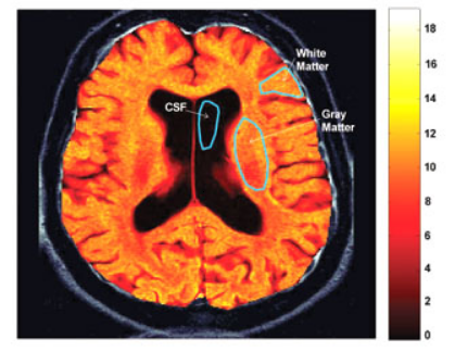
Structural images, which are obtained by MRI scanners, are weighted images that are called T1 and T2 (which are short for T1-Weighted & T2-Weighted). In Relaxometry analysis the real values (unweighted) are obtained using consequential measurements and equations that govern these procedures.
Since these relaxation values are inherent to the tissue, this technic is used to evaluate the true nature of the tissue (where instead of contrast, inherent characteristics of the tissue are important).
This method is capable of identifying the changes in the natural characteristics of the tissue, in the nervous system caused by pathologic changes.
The main use of this technique is to study the chemical solutions and contrast agents.
This method also provides the ability to check the contrast of agents within the animal body.
The effects of a substance in the T1 & T2 Relaxation values can be measured by this method.


Related Posts