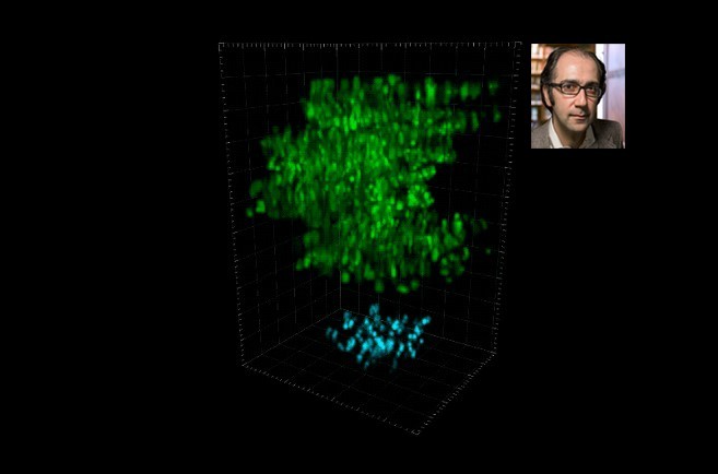
New microscopy technique peers deep into the brain
In order to understand the brain, scientists must be able to see the brain—cell by cell, and moment by moment.
In order to understand the brain, scientists must be able to see the brain—cell by cell, and moment by moment. However, because brains comprise billions of microscopic moving parts, faithfully recording their activity comes with many challenges. In dense mammalian brains, for example, it is difficult to track rapid cellular changes across multiple brain structures—particularly when those structures are located deep within the brain.
A novel microscopy technique, developed by Rockefeller scientists, integrates new and existing approaches to help build a more cohesive picture of the brain. Described in Cell, the technology captures cellular activity across large volumes of neural tissue, with impressive speed and at new depths.


Related Posts