Brain Connectivity Patterns on fMRI Differ in Chronic and Episodic Migraine
Abnormal, time-varying brain connectivity between the thalamo-cortical and frontoparietal pathways may contribute to symptoms of migraine. These findings from an analysis of functional magnetic resonance imaging (fMRI) data were published in The Journal of Headache and Pain.
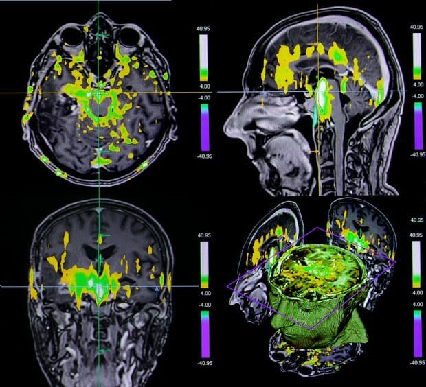
Study researchers recruited patients with episodic or chronic migraine (n=20) and participants in a healthy control group (n=26) by local advertisement. All participants underwent fMRI assessment at the University of Michigan in which they were asked to relax and keep visual focus on a screen and try not to think of anything. Blood-oxygen-level-dependent signal variability (BOLDSV) was calculated across low frequencies (0.01-0.198 Hz).
Patients and control group participants were well balanced for age (P =.428) and sex (P =.818). Among patients, those with chronic or episodic migraine did not differ for disease duration (P =.945), ictal thermal pain threshold (P =.202), ictal pain intensity (P =.246), or the 6-item Headache Impact Test (P =.086).
Specific regional variation in BOLDSV patterns were observed between control group participants and patients with migraine, and between patients with chronic migraine and those with episodic migraine. Study researchers averaged these observations, and cross-correlation graphs indicated BOLD signal fluctuations were in a temporally synchronized pattern between the trigeminal spinal-thalamo-cortical and the frontoparietal pathways.
Using a direct conditional correlation analysis, patterns of strength (P <.05) and variability (P <.05) between ventral posteromedial nucleus and primary somatosensory cortex with dynamic functional connectivity (dFC) differed between control group participants and patients. Patients with chronic migraine had significantly greater dFC strength and lower variability compared with control group participants (all P <.05). Patients with episodic migraine had higher variability of dFC (P <.05).
All patients had decreased strength of dFC between inferior parietal cortex and right dorsolateral prefrontal cortex compared with control group participants (P <.01), more driven by patients with episodic (P <.01) than chronic (P <.05) migraine.
Headache severity was correlated with increased BOLDSV in the left medial pulvinar nucleus (r, 0.697; P =.006), primary somatosensory cortex (r, 0.662; P =.010), right spinal trigeminal nucleus (r, 0.595; P =.025), and dorsal posterior insula (r, 0.558; P =.038).
This study was limited by its low sample sizes of patients with chronic and episodic migraine and because generalized findings among all patients may be driven by only a subset of individuals.
Study researchers concluded, “Migraine is associated with alterations in temporal signal variability in the ascending trigeminal somatosensory and top-down modulatory pathways, which may explain migraine-related pain and allodynia. Contrasting patterns of time-varying connectivity within the thalamo-cortical and frontoparietal pathways could be linked to abnormal network integrity and instability for pain transmission and modulation.”
https://www.neurologyadvisor.com/topics/migraine-and-headache/brain-connectivity-patterns-on-fmri-differ-in-chronic-and-episodic-migraine/


Related Posts