

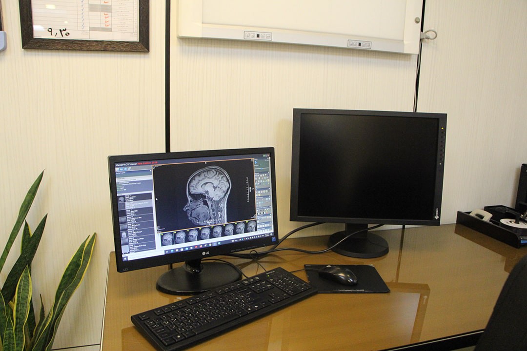
Magnetic Resonance Imaging (MRI)
Magnetic Resonance Imaging (MRI) is a noninvasive medical imaging technique which can be used by physicians or researchers for many purposes including diagnosis of the diseases, staging of them and monitoring their progression, evaluation the responses to therapeutic interventions, and drug discovery. An MRI scan uses a strong static magnetic field, a gradient magnetic field, radio frequency pulses and processing devices to generate detailed pictures of body structures. Any part of body such as brain, spinal cord, etc. can be examined by this imaging technology.
List of Equipment(Top)
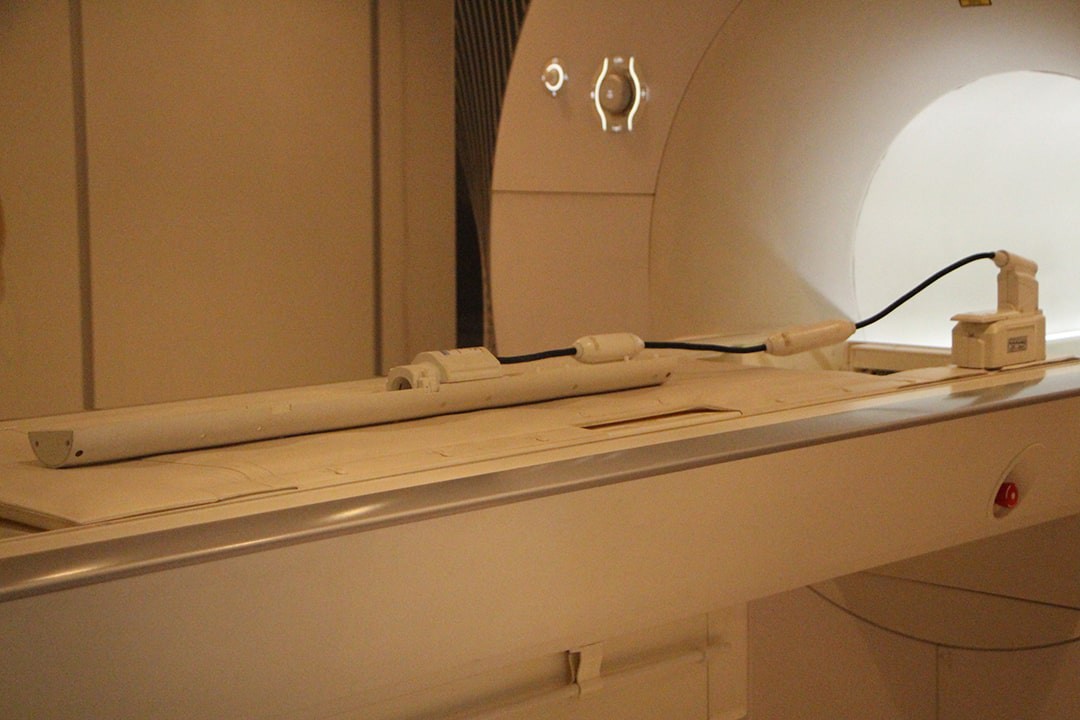
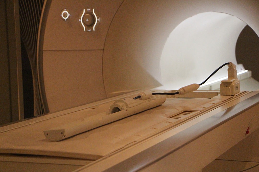
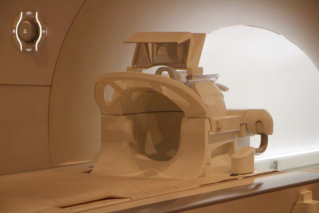
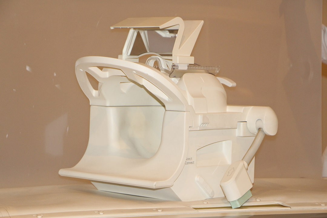
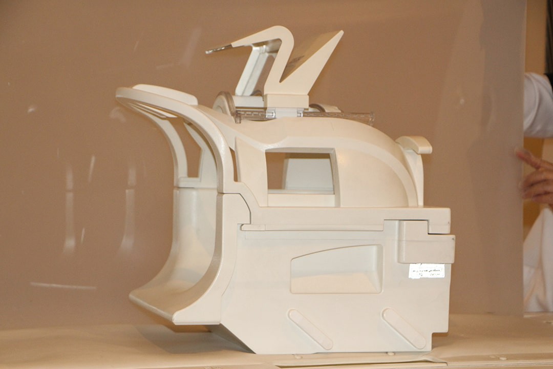
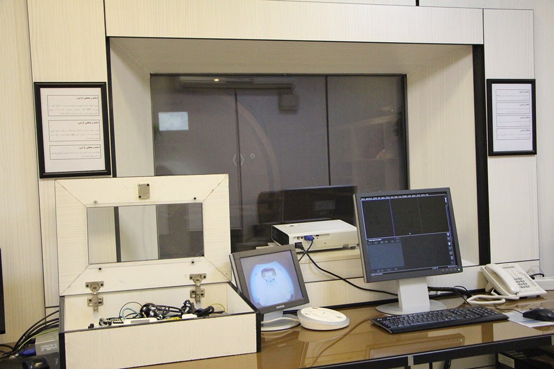
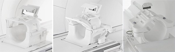
MR Scanner(Top)
- Equipped with a 3.0 Tesla Siemens Prisma MRI Scanner
- High homogeneity across its large 50 cm FOV
- State-of-the-art zero helium boil-off technology
- Up to 204 simultaneously connected coil elements in combination with the fully integrated 64 independent RF channels
- Tim 4G coil technology with Dual Density Signal Transfer and DirectConnect Technolgy

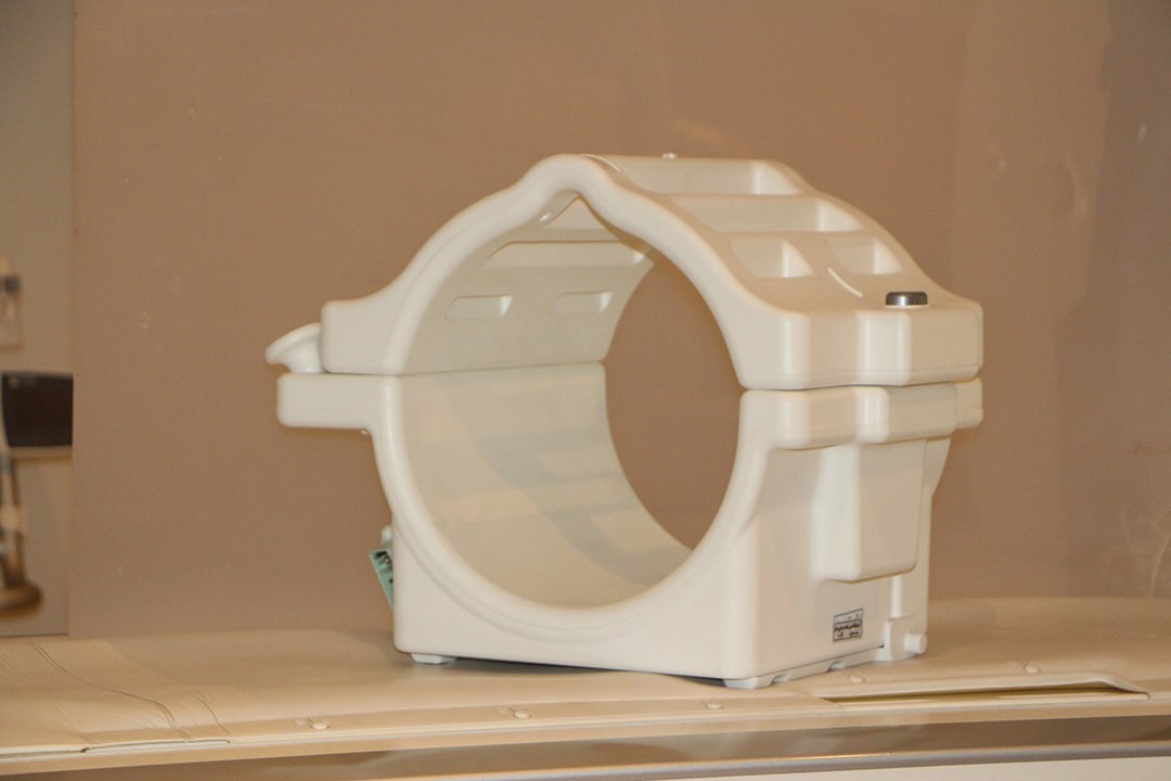
Coils(Top)
- 20 channel head/neck coil
- 64 channel head/neck coil
- Transmit/receive CP head Coil
- 4 channel large and small flex coils
- 18 channel body flex coil
- 31P 1H volume head coil
- 13C 1H volume head coil
- 1H Rat Coil (With rat holder)
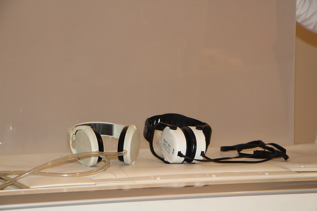
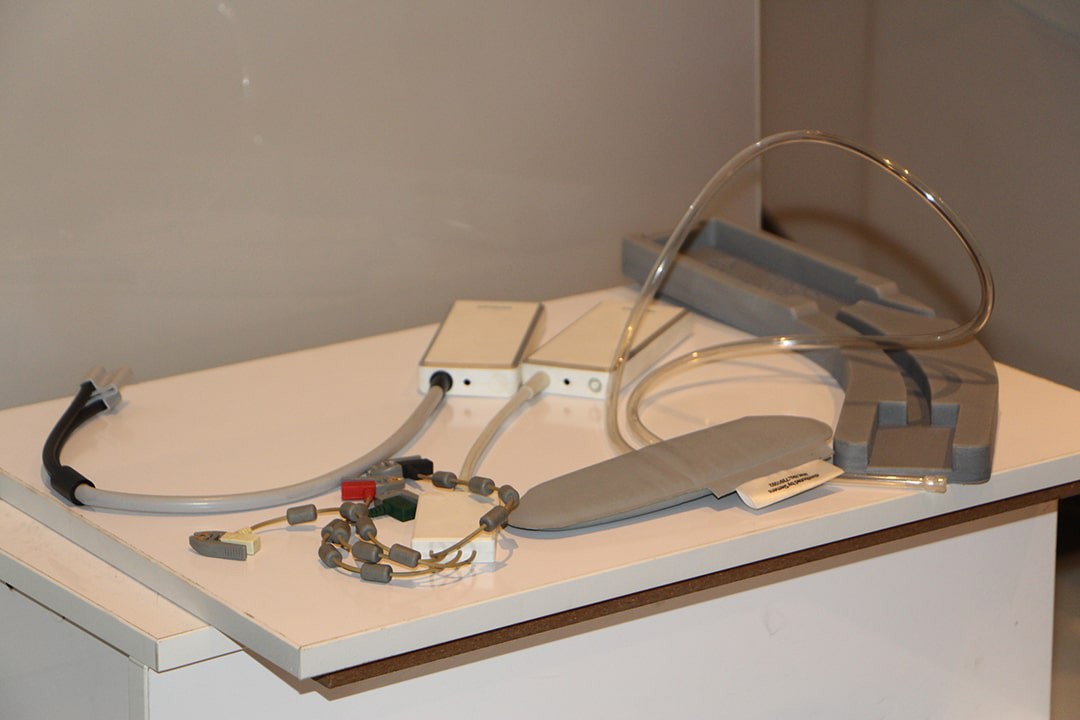
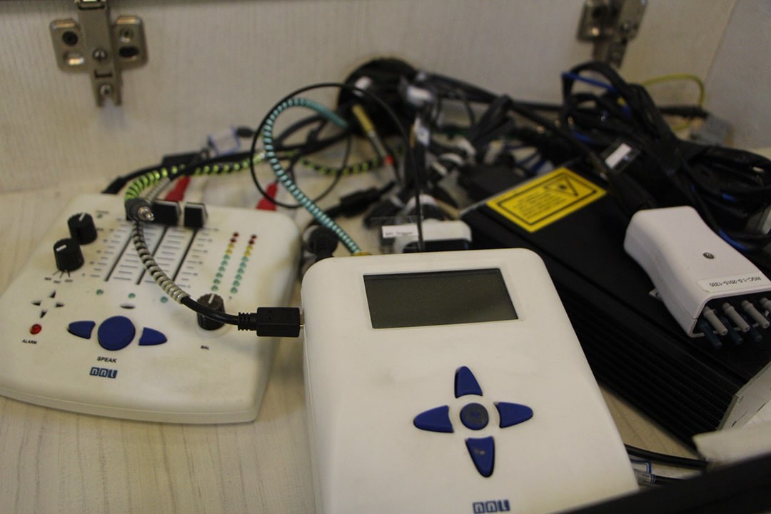
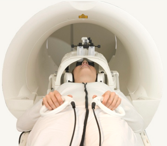
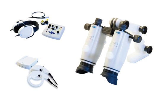
Equipment for fMRI(Top)
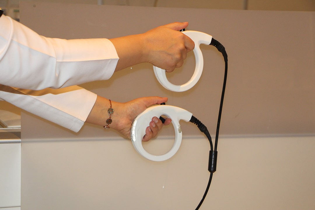
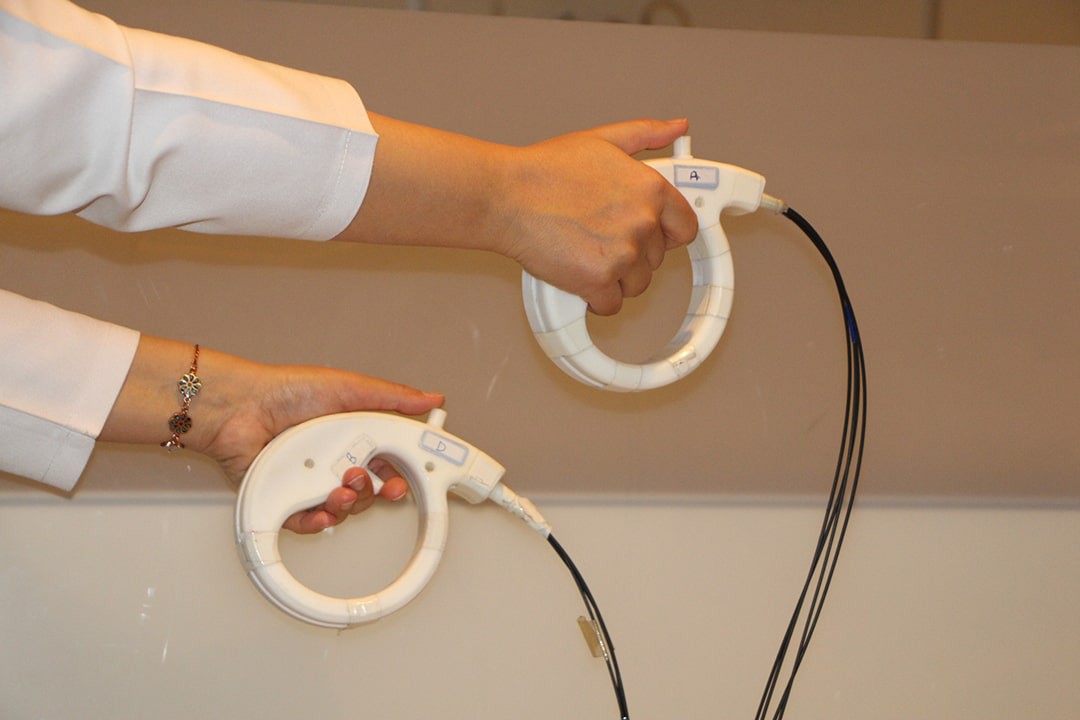
- Response Grips
- Coil Mounted Display
- Eye Tracking Camera
- Audio System
- Ear Plug
- Communication Console
- Sync Box
Physiological Imaging(Top)
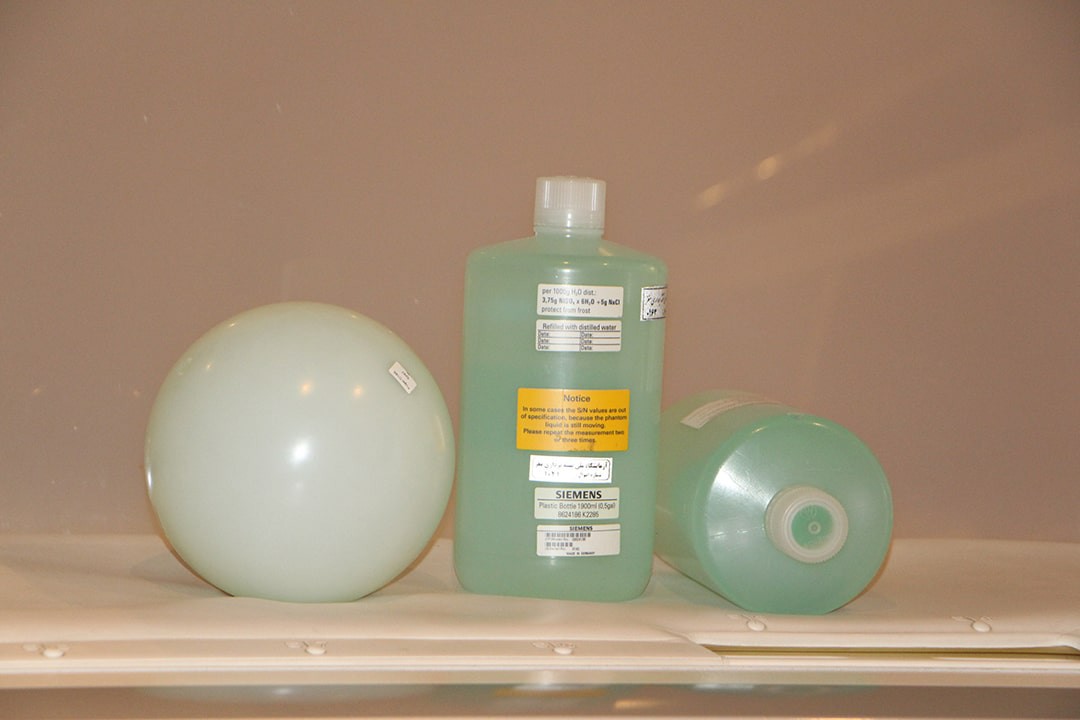
Physiological Measurement Unit (PMU)
- PERU (Physiological ECG and Respiratory Unit): ECG and Respiratory
- PPU (Peripheral pulse unit): Pulse sensor
- External trigger input

More Equipments(Top)
- 50cm FOV, with the industry best homogeneity
- Whole-body, superconductive Zero Helium Boil-Off 3T magnet
- XR 80/200 gradient engine generates
- Head/Neck 20 DirectConnect
- Spine 32 DirectConnect
- Body 18, Flex Large/Small 4
- syngo MR software
- State of the art host computer
- Tim Table #P
- Flow Quantification
- Argus Flow
- Neuro Perfusion Package #T+D
- Diffusion Tensor Imaging #P
- Inline BOLD Imaging #TimInline technology
- Arterial Spin Labeling (ASL)
- SWI #Tim (Susceptibility Weighted Imaging)
- fMRI Trigger Converter
- Single Voxel Spectroscopy #Tim
- 2D Chemical Shift Imaging #Tim
- 3D Chemical Shift Imaging #Tim
- Spectroscopy Evaluation syngo
- Head/Neck 64 #P,Sk
- Patient Supervision TV #T+D
- 31p 1H volume head coil
- 13C – 1H volume head coil
- 1H phased array for rat brain
- Animal Holder for rats
- Tx/Rx CP Head Coil #Sk
List of services(Top)
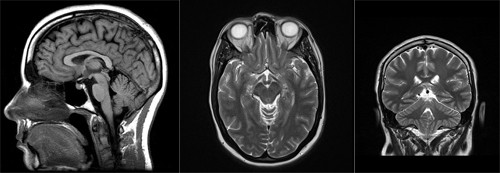
Anatomical Brain Imaging(Top)
Magnetic resonance imaging (MRI) is an imaging technique used primarily in medical settings to produce high quality images of the soft tissues of the human body.
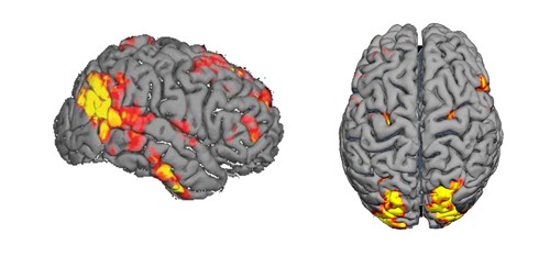
Functional MRI (fMRI)(Top)
fMRI is a worldwide diagnostic tool for learning how a normal, diseased or injured brain is working, as well as for assessing the potential risks of surgery or other invasive treatments of the brain and how different parts of the brain respond to external stimuli or passive activity in a resting state.
Common services of fMRI:
- Study the anatomy of the brain
- Determine which part of the brain is handling critical functions such as thought, speech, movement and sensation.
- Help to assess the effects of stroke, trauma or neurodegenerative diseases such as Parkinson Disease on the brain function.
- Monitor and stage the growth and function of brain tumors.
- Guide the planning of surgery, radiation therapy, or other surgical treatments for the brain.
Diffusion-Weighted Magnetic Resonance Imaging(Top)
This imaging method uses the diffusion of water molecules to generate contrast in MR images and allows the mapping of the diffusion process of molecules, mainly water, in biological tissues, in vivo and non-invasively.
Applications:
- DWI is currently a standard in diagnosis of ischemic stroke
- Diagnosis of epilepsy, neurotoxicity etc.
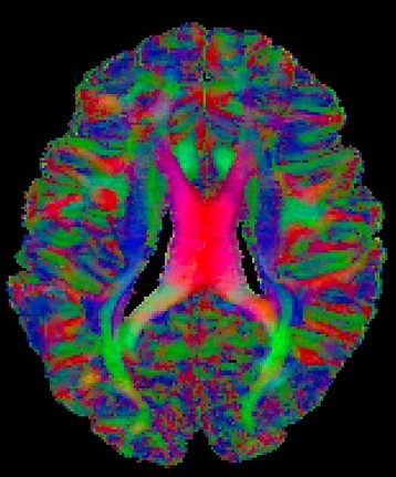
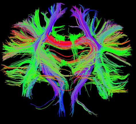
Diffusion Tensor Imaging(Top)
DTI relies on estimating diffusion tensors, which can be obtained through taking DW images in multiple directions.
Through DTI of gray matter, connectivity in brain could be understood better
Diffusion Tensor Imaging is a non-invasive way of understanding brain structural connectivity and macroscopic axonal organization
Diffusion MR Images can measure water proton displacements at the cellular level and probing motion at microscopic scale (mm), orders of magnitude smaller than macroscopic MR resolution (mm)
It provides accurate estimation of the tensor field of the wight matter in the brain and accurate estimation of the connectivity between different brain regions is of great clinical and research interest
Applications:
- Advanced knowledge on anatomy of brain
- Diagnosis of white matter and gray matter related pathologies like lesions, axonal injuries, demyelination, multiple sclerosis
- Psychiatric disorders like Schizophrenia, ADHD, neurological and neuro-degenerative disorders like cognitive impairment, Alzheimers’ disease etc
- Parcellation and connectivity mapping in brain, in the Human Connectome Project, which aims at mapping the brain at regional level
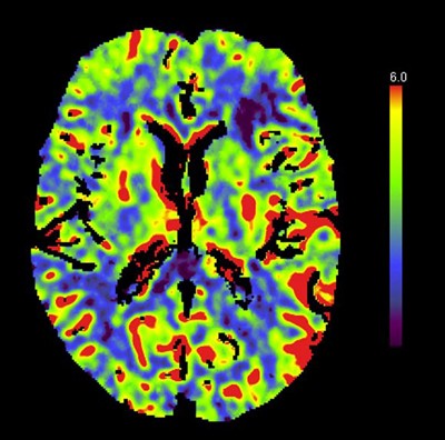
Perfusion Imaging(Top)
Perfusion is one of the most important physiologic and pathophysiologic parameters and can be assessed non-invasively with MRI.
It refers to the delivery of blood to the cells via capillaries in a certain time period and is identified with blood flow which is measured in milliliters per minute per 100g of tissue
Applications:
- With the ability to ascertain data on the blood flow to vital organs such as the brain, accurate choices on treatment for patients could be made
- To trace a number of neurological illnesses to abnormal blood flow such as ischemic stroke, cerebral neoplasia, Alzheimer, MS, brain tumors
- To assess patients with neurodegenerative diseases
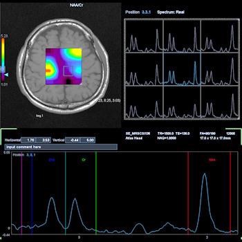
Magnetic Resonance Spectroscopy(Top)
Magnetic resonance spectroscopy is a non-invasive analytical technique that used to measure the levels of different metabolites in body tissues.
The MR signal produces a spectrum of resonances that corresponds to different molecular arrangements of the isotope being "excited". This signature is used to diagnose certain metabolic disorders, especially those affecting the brain and to provide information on tumor metabolism.
Applications:
- Brain tumors
- Dementia and movement disorders such as Alzheimer's disease, front temporal dementia, motor neuron disease, Parkinson disease/Parkinsonian syndromes etc
- Head trauma
- seizure disorders
- depression
- Psychiatric disorders (e.g., attention-deficit/hyperactivity disorder, autism spectrum disorders, bipolar disorder, depression, emotional dysregulation, obsessive-compulsive disorder, and schizophrenia)
Some Useful Links(Top)
- Dr. Alexander Leemans, Image Sciences Institute, University Medical Center Utrecht
- Dr. Peter Kochunov, Maryland Psychiatric Research Center, University of Maryland
- Professor Paul Thompson, University of Southern California1000 Functional Connectomes Project
Experts in this field