پردازش و آنالیز تصاویر
معرفی آزمایشگاه پردازش تصویر
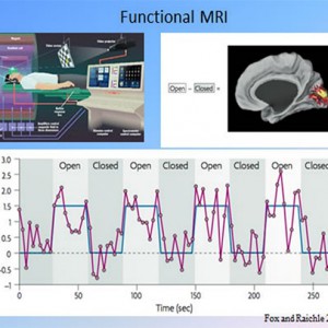
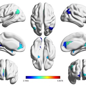
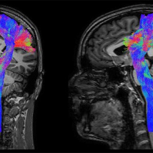
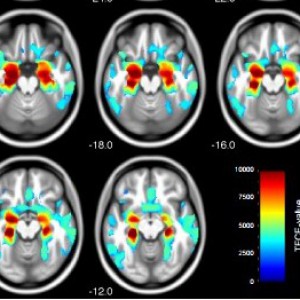
مغز انسان به عنوان پیچیده ترین سیستم شناخته شده، الهام بخش علوم مختلف بوده است. به طوری که در بسیاری از علوم، ساختار یا کارکرد مغز انسان، الهام بخش توسعه علوم دیگر بوده است. در حال حاضر بهترین راه شناخت ساختار و عملکرد مغز، استفاده از تصاویر تشدید مغناطیسی (MRI) است. تصاویر MRI به صورت خام حاوی اطلاعات خاصی نیستند. به عنوان مثال دیدن تصاویر fMRI به صورت خام حتی برای یک پزشک متخصص نیز مفهومی ندارد. بنابراین استخراج یک تصویر قابل بررسی در مدالیته های پیشرفته MRI مانند fMRI و DWI نیازمند انجام مجموعهای از پردازش های اولیه و ثانویه روی این داده ها است. از طرفی انجام برخی از این پردازش ها نیازمند دانش حداقلی در زمینه پردازش سیگنال و تصویر است که تمامی پژوهشگران در علوم مختلف آشنایی تخصصی با این حوزه ندارند.
آنچه ما در آزمایشگاه پردازش تصویر در اختیار پژوهشگران می گذاریم، دانش و تجربه ی پردازش تصاویر مغز است تا پژوهشگران علوم مختلف بی نیاز از درگیری در مسائل پیچیده پردازش تصویر، بر حوزه تخصصی خود تمرکز کرده و پژوهش خود را پیش ببرند. نتایج قابل تفسیر، کمی و قابل اطمینان از طریق روال های آنالیز قابل اطمینان و متداول و به صورت تخصصی متناسب با نیاز پژوهشگران در اختیارشان قرار داده می شود.
تصویر برداری تشدید مغناطیسی (MRI) و قابلیت های آن
قبل از شروع یک مطالعه ی Neuroimaging لازم است درک صحیحی از قابلیت های MRI داشته باشیم تا متناسب با این قابلیت ها، علاوه بر استفاده از حداکثر ظرفیت این ابزار، از انتظارات غیرواقعی وگمراه کننده در این حوزه جلوگیری کنیم.
به طور کلی می توان تصاویر MRI را به دو دسته تقسیم بندی کرد:
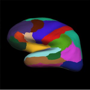
- تصاویر ساختاری
- تصاویر عملکردی
هر کدام از این تصویربرداری ها به چند مدالیتی تقسیم می شوند که بر اساس نوع اخذ داده میتوانند کنتراست خاصی را در اختیار محقق قرار دهند. به عبارتی میتوان MRI را به دوربینی تشبیه کرد که با تنظیمات مختلف از پدیده ها یا ویژگی های متفاوتی تصویر تهیه می کند. کنتراست در تصاویر تشدید مغناطیسی (که باعث شکل گیری تصویر ظاهری یک ساختار برای ما میشود)، بستگی به نحوه ثبت آن تصویر دارد. می توان با تنظیم پالسهای فرکانس رادیویی و گرادیانها و انتخاب دقیق زمان بندی آنها، از ویژگیهای مختلف بافت تصویربرداری کرد.
کارشناسان این بخش