Simple Magnetic Resonance Imaging (MRI) enables us just to see anatomical changes of a lesion. However, the process of growth of a lesion is in the form of biological, physiological and anatomical changes, respectively. So we need the methods that can show us the changes and leveling of a lesion in the early stages. Perfusion is the passage of fluid through the circulatory system or lymphatic system to an organ or a tissue, usually referring to the delivery of blood to a capillary bed in tissue.
Clinical applications of Perfusion imaging:
- Identifying areas with tumors
- Tumor classification
- Identifying the difference between a benign and a malignant tumor
- Grading all kinds of malignancies
- Diagnosing of necrotic tissue by radiation
- Checking the response to treatment
- Identifying other disorders in the brain
There are different approaches based on exogenous or endogenous contrast agent which measure cerebral perfusion with MRI:
- Dynamic susceptibility contrast-enhanced (DSC)
- Dynamic contrast-enhanced (DCE)
- Arterial spin labeling (ASL)
In the first two methods, we use exogenous contrast agent to have more sensitivity and spacial contrast. But ASL uses magnetically labeled arterial blood water protons as an endogenous tracer.
ASL: Arterial spin labeling (ASL) is a non-ionizing and completely non-invasive MRI technique for measuring tissue perfusion (blood flow), which uses magnetically labeled arterial blood water protons as an endogenous tracer. These benefits make ASL very suitable for perfusion studies in healthy individuals, patients with renal insufficiency and those who need repetitive follow-ups.
We can measure the cerebral blood flow (CBF) by ASL. A CBF image is computed by applying a set of measured or assumed physiological and MR parameters on the ASL-signal image to obtain voxelwise flow values in absolute physiological units of flow. CBF represents the amount of blood that passes through the tissue per unit of time. The unit is mL/100g/min.
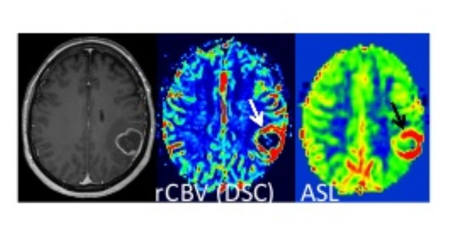
Figure 1: An example of perfusion image to identify areas with tumors
List of services:
The ASL data is analyzed using SPM at NBML image processing laboratory.