
Functional Near-infrared Spectroscopy (fNIRS) Section
Functional Near-infrared Spectroscopy (fNIRS) is a functional optical imaging technology that measures neural activity and hemodynamic responses in the brain. It measures NIR light absorbance of hemoglobin in the blood with and without oxygen and provides information about ongoing brain activity similar to functional MRI studies. In this method, a ray of light is used near the visible spectrum of light is emitted at the scalp. This light will be multiply scattered and partly absorbed. By measuring the changes of the characteristics of this light wave (specially the absorption spectra of light absorbing molecules) hemodynamic responses are evaluated. In other words, fNIR technology measures relative changes in the concentration of oxygenated hemoglobin (oxyHb), deoxygenated hemoglobin (deoxyHb), and total hemoglobin (oxyHb+deoxyHb) for real-time and continuous monitoring of brain dynamics. Since fNIR and fMRI are both sensitive to similar physiologic changes, they are comparative methods. fNIR has several advantages over fMRI in terms of cost and portability; but cannot be used to measure cortical activity more than 4 cm deep due to limitations in light emitter power and has more limited spatial resolution.
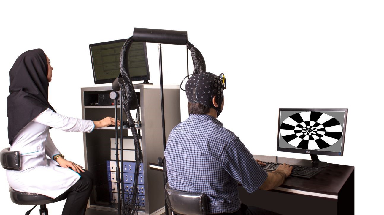
The fNIR system can also be readily integrated with other physiological and neurobehavioral measures including eye tracking, pupil reflex, respiration, blood pressure, ECG, and electrodermal activity to comprehensively evaluate human brain activity. fNIR system allows researchers to quantitatively analyze brain functions such as working memory, decision making, planning, attention, problem solving, and etc while subjects performing cognitive tasks.
The technology empowers researchers by providing greater flexibility for real-life study design, including working within complex lab environments and deploying in various field settings such as operating room, classroom, cockpit, control stations, training simulators, and etc.
Advantages vs. limitations
The advantages of fNIR are as follows:(Top)
- It is a non-invasive technique without exposure to ionization radiation, making it safe for subjects.
- Wireless and wired solutions are available for adult and pediatric subjects, making it convenient for all ranged subjects.
- It is relatively inexpensive compared to MRI or PET scans.
- It is portable, small, and relatively easy to set-up. Subjects are less physically constrained than for fMRI, PET, and MEG.
- It can measure hemodynamic signals with a temporal resolution of 100 Hz or higher, significantly greater than fMRI or PET.
- It can be used while subjects are performing variety of tasks.
- It can be used for populations that may not be able to readily tolerate fMRI magnet (e.g., schizophrenics, autistic children, neonates)
- It is able to monitor cerebral oxygenation for a long duration (e.g., premature and other high-risk infants).
However the limitations of this technology include:(Top)
- It cannot be used to measure cortical activity more than 4 cm deep due to limitations in light emitter power.
- It has more limited spatial resolution compared to MRI.
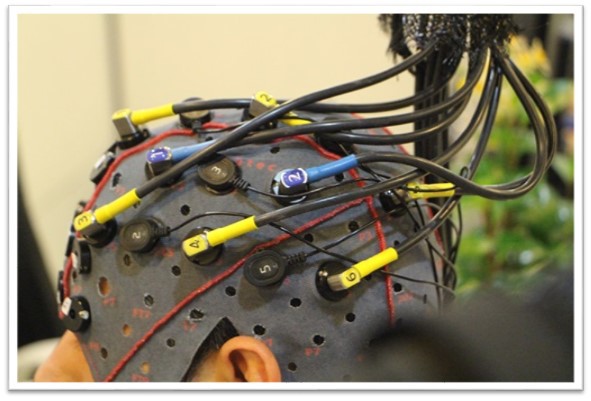
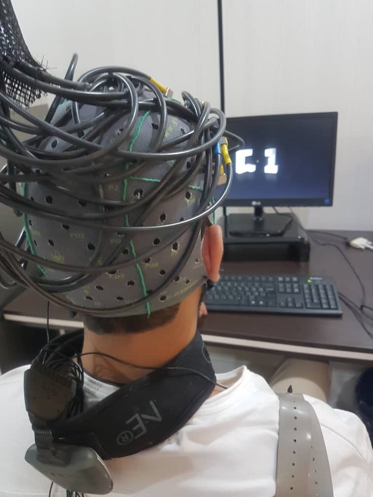
- Similar to EEG, care must be taken to ensure that hair does not severely block the transmission of light between the source and detector and the scalp.
List of Services(Top)
- Brain Oxygenation monitoring
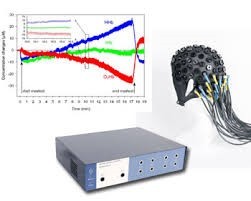
- Measures oxy-, deoxy-, and total hemoglobin concentration changes and optionally absolute concentrations and tissue saturation index(TSI).
- Quantitative analysis of brain functions
- Concurrent fNIRS/MRI
- Concurrent fNIRS/ other modalities
List of Equipment(Top)
What do we have in fNIRS Section of NBML?
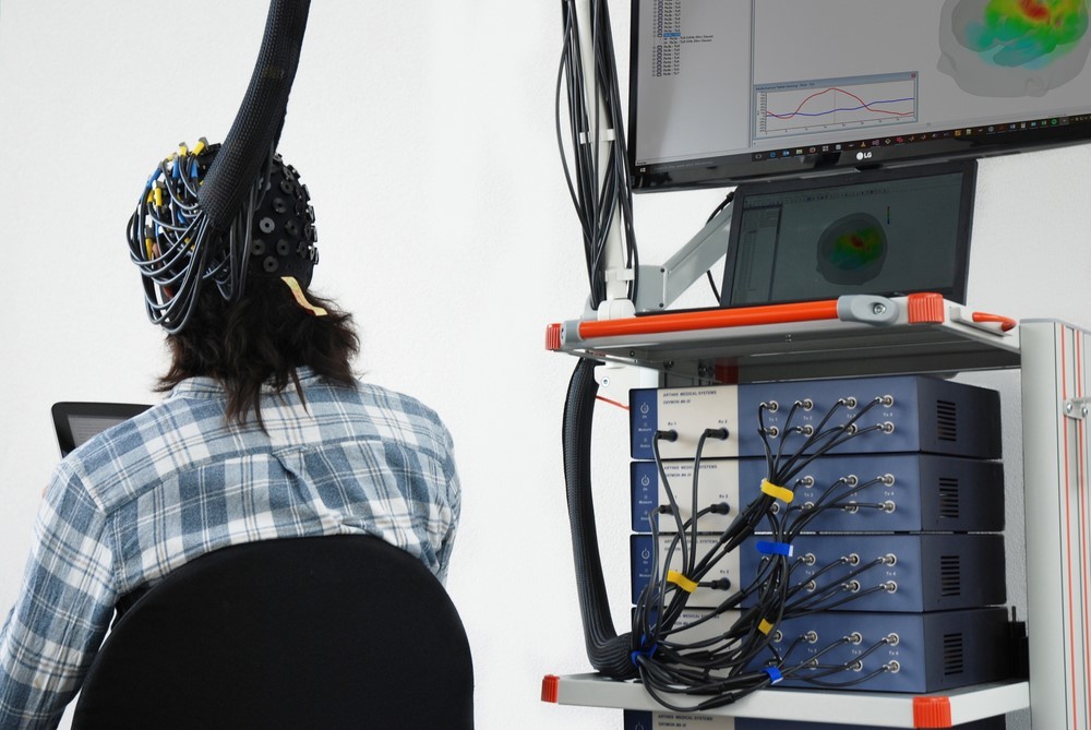
This section is equipped with a 48 channel MR compatible OxyMonfNIRS from Artinis. The MR compatible fNIR system empowers researchers by providing high spatiotemporal resolution image of brain activities. This is a highly advanced research tool with the main specifications as follows:
- Measures oxy-, deoxy-, and total hemoglobin concentration changes and optionally absolute concentrations and tissue saturation index.
- Fast data collection at 50 Hz as standard – up to 250 Hz is possible.
- Sampling time Noise: From 0.1 Hz to 50 Hz (up to 250 Hz optional)
- ~0.001 standard deviation in optical density at a total of ~6 optical densities at 10Hz measurement frequency
- Standard configuration with two wavelengths per channel
- Interference: With NMR compatible fibers the instrument can be used inside the
- MRI. EEG/ECG does not interfere the optical signal
Specifications / Main Items:
- Four units, each consisting of four transmitters (Tx) modules and two light receiving (Rx) module
- Win 7 Desktop computer with software
- Application software
- Fibers for standard setup (2×24 Channel)
- Calibration tool
- Headcap without holders
- Spacers for the headcap with 3 cm equidistant markings
- NMR fiber set
- AD input box
- Computer interface for psychology research
- Remote event generator
- Real time data export to Matlab in progress
- Offline data export to Matlab
- Data conversion for SPM software packages in Matlab
Experts in this field