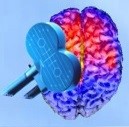
Transcranial Magnetic Stimulation (TMS)
What is TMS?
Faraday discovered electromagnetic induction in 1831, About 60 years later, Arsenne d'Arsonval detected phosphene and vertigo in effect of electromagnetic induction. In some persons, "syncope" was detected when the subject’s head was placed inside an induction coil. Transcranial magnetic stimulation has been first successfully used in 1985 by Anthony Barker, Reza Jalinous at the Royal Hallamshire Hospital in Sheffield, England. Since then TMS has been employed in various research and clinical (diagnostic and therapeutic) applications. This device works based on Faraday’s law of electromagnetic induction so that a large electrical current passes through TMS coil during a very short time and this induces very small electrical currents in the cerebral cortex. The coil is connected to a stimulator which generates the electric current passing from the coil.
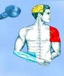
TMS Magnetic field, which is generated by the coil, passes through high-resistance structures such as bone, fat and skin without harming the body. Furthermore, it induces an electric field, which depolarizes or hyperpolarizes neurons in the underlying tissue.
TMS pulses can be applied as “single”, “paired”, or “train” (‘‘conventional” or ‘‘patterned”) and each form can be used for special application. Single-pulse TMS can be used in cortical mapping, causal chronometry of brain-behavior relations, central motor conduction time studies, etc. Paired pulses can be used to study intracortical facilitation and inhibition, and cortico–cortical interactions. Magnetic pulses can also be paired with a peripheral electric pulse called paired associative stimulation (PAS). Trains of pulses can generate fascinating effects depending on their frequency and pattern, such as lasting inhibitory or facilitatory aftereffects. This protocol has also been used in brain mapping studies to establish causality between brain and behavior. As a brain mapping technique with good spatial (in the order of few millimeters) and temporal (in the order of a few tens of milliseconds) resolution, TMS can be used to reveal the location and sequence of brain activities. TMS is an FDA (Food and Drug Administration) approved intervention for major depression. It can be also used for treatment of migraine, and for presurgical motor and language mapping.
What do we have in TMS section of NBML?
Three main TMS applications:
 |
 |
 |
| Clinical Diagnostic & Brain Research | Therapy Young market |
Rehabilitation |
| Transcranial Magnetic stimulation (TMS) and Motor Evoked Potential (MEP) | Transcranial Magnetic stimulation (TMS) | functional Magnetic Stimulation(fMS) |
|
|
|
| Customer Group | Customer Group | Customer Group |
|
|
|
| Applications | Applications | Applications |
|
|
|
List of Equipment (Top)
Main items in TMS section are as follows:
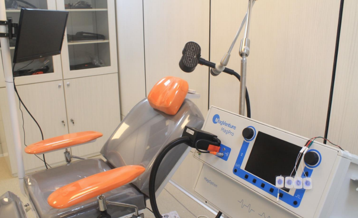
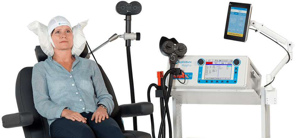
1. MagPro X100 incl. MagOption
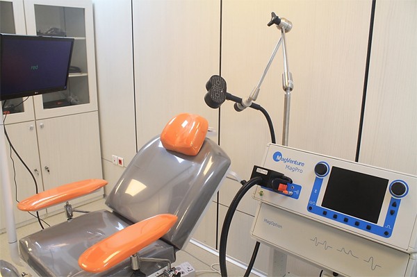
Figure 1: TMS equipment in NBML lab
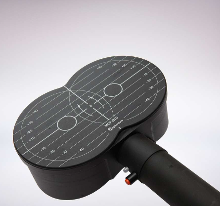
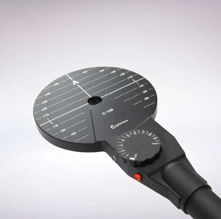
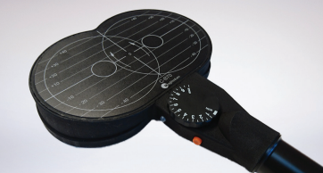
2. Coils:
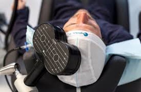
Figure 2: TMS coil configuration
- Coil C-100
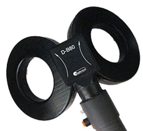
- Coil D-B80
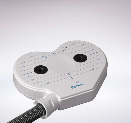
- Coil MRi-B91
- Coil MCF-B65
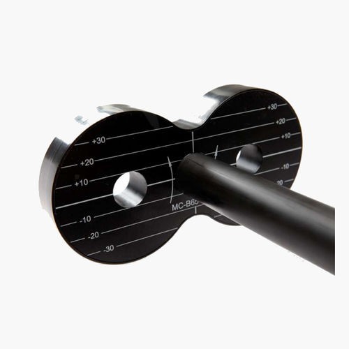
- Coil MC-B65-HO
- Coil Cool-B65 A/P
- Coil Cool-40(rat coil)
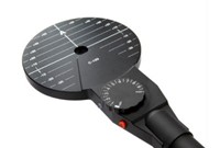
| Circular Coils A “general purpose coil” which can be positioned over many parts of the body to stimulate a fairly large area. | ||
| Model | ||
| C-100* | Weight of transducer head: 0.6 kg Cable length: 1.7 m Dimensions of transducer head(WxLxH): ø123 x 11.5 mm Inner diameter: 20mm Outer diameter:110mm Winding height: 6mm Number of windings: 14 Number of stimulations, before warm-up at ambient temperature 20°C: 400 pulses Mean output 75% of maximum at 1pps. |
 |
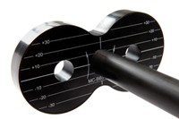
| MC-B65-HO | Dimensions of transducer head: 165 x 85 x 22mm Inner diameter 35mm Outer diameter 75mm Winding height 11mm Number of windings 2x (2 x 5) Orthogonal handle Pulses before warmup: 350 Compatible with R20, R30, R100 & X100 Usable with MagPro Compact w/ converter |
 |
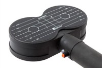
| MCF-B65 | Dimensions of transducer head 172 x 92 x 53 mm Weight of transducer head 1.5kg Cable length 2m Inner diameter 35mm Outer diameter 75mm Winding height 12mm Number of windings 2x (2 x 5) Pulses before warmup: 2000 Trigger button Cooled with static fluid Compatible with R20, R30, R100 & X100 |
 |
| Cool-B65 A/P | Weight of transducer head 3 kg Cable length 2m Dimensions of transducer head 172 x 92 x 80 mm Inner diameter 35mm Outer diameter 75mm Winding height 12mm Number of windings 2x (2 x 5) Pulses before warmup: >20.000 Cooled dynamically with external coil cooler unit Compatible with R30 & X100 Works both as active and placebo. Excellent for clinical trials Designed for advanced double-blinded clinical studies requiring a very high number of stimuli. |
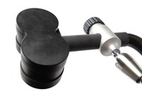 |
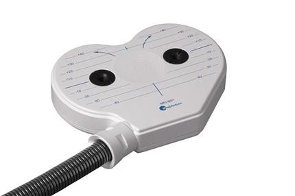
| Cool D-B80 | Weight of transducer head 1.8 kg Cable length 2 m Dimensions of transducer head: 2 x ø110mm Thickness: 30mm Angle: 120º Pulses before warmup: 500 Trigger button Compatible with R20, R30, R100 & X100 Usable with MagPro Compact w/ converter |
 |
| Special Coils We have developed a number of special coils for our various system solutions. Custom designed coils are available as well as modifications to existing coils. | ||
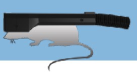
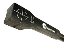
| Cool-40 Rat Coil | Weight of transducer head: 0.5 kg Cable length: 1.4 m Dimensions of transducer head: 52 x 54 x 42 mm Diameter: ø40 mm Small focal coil for use on rodents Allows a high number of stimuli Can be used inside a PET and SPECT scanner Cooled with the High-Performance Cooling System Not for human use |
 |
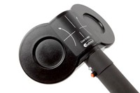
| MRi-B91 | Weight of transducer head: 1.1 kg Dimensions of transducer head: 175 x 142 x 30 mm Cable length: 6 m Inner diameter: 2 x ø35/ø52 Outer diameter: 2 x ø79/ø92 Winding height: 16 mm Number of windings: 2x (2x4) Distance between centers: 79 mm Temperature warm-up at 100%: +30m°C/pulse Compatible with R30 & X100 For use in MRi scanners up to 4 Tesla Used with TMS-fMRi System Solution which includes remote control, power line filter & emergency stop |
 |
3. TMS Sham Noise Generator can produce TMS noise for sham studies
4. MagProbe (DIN connector)
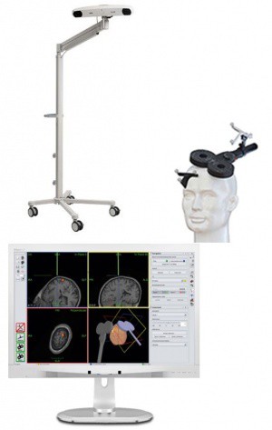
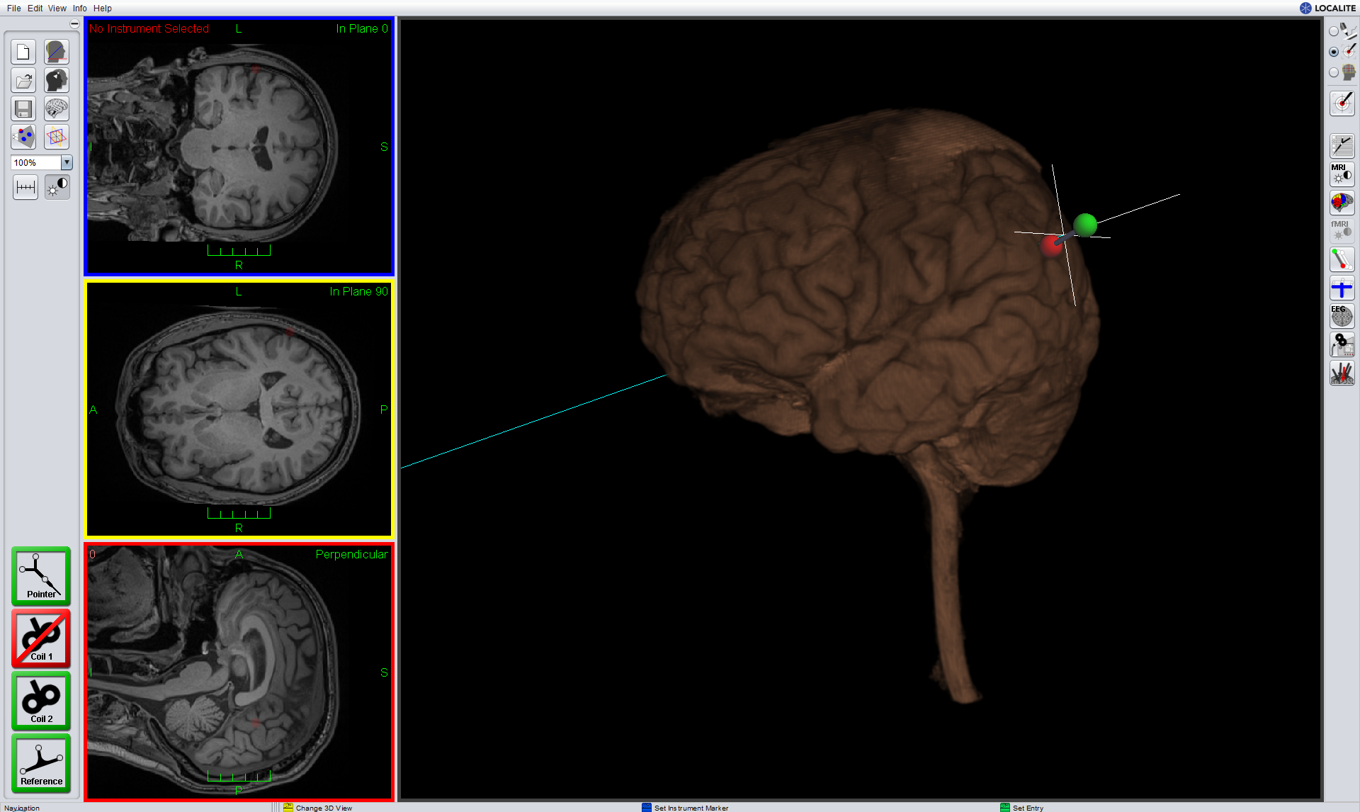
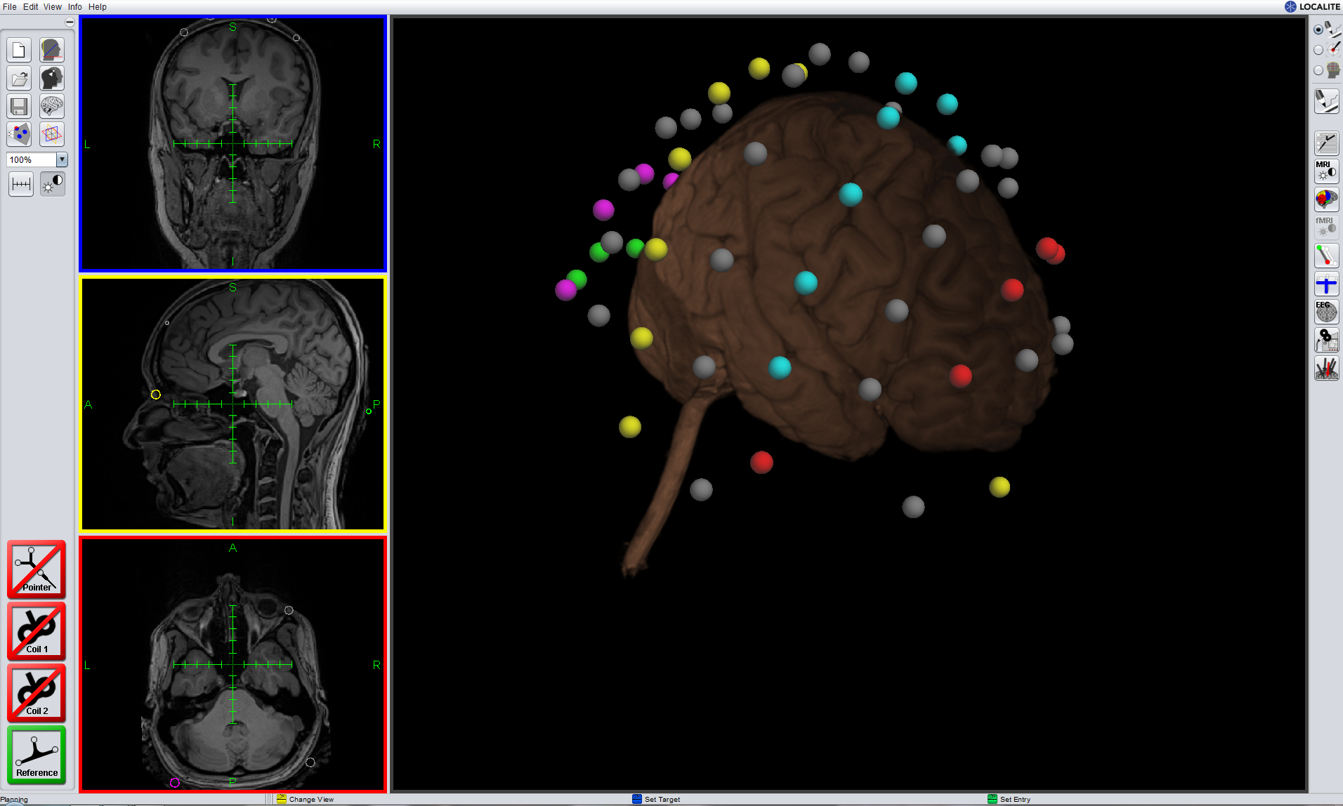
5. TMS Navigator from localite (MR-based and MR-less)
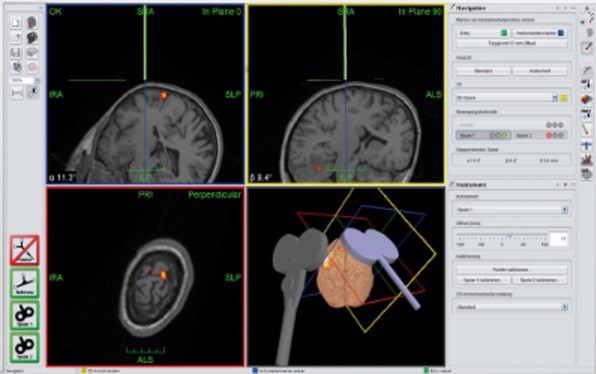
Figure 3: MR-Based System, using individual MRI data
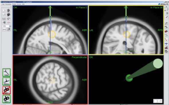
Figure 4: MR-Less Navigation System supports navigated stimulation using MNI atlas, without MRI
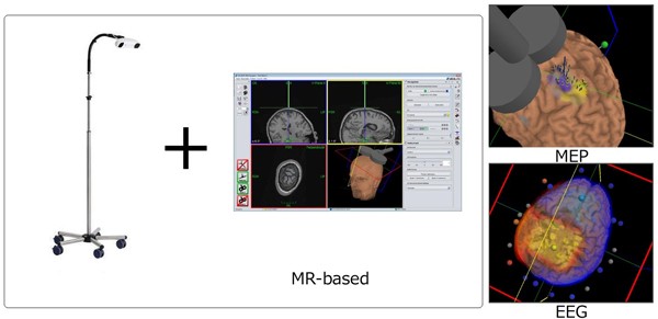
Figure 5: Navigation camera and MEP and EEG projection on the brain in Navigation system
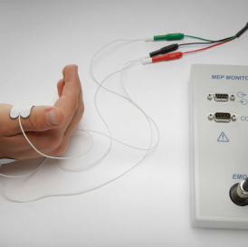
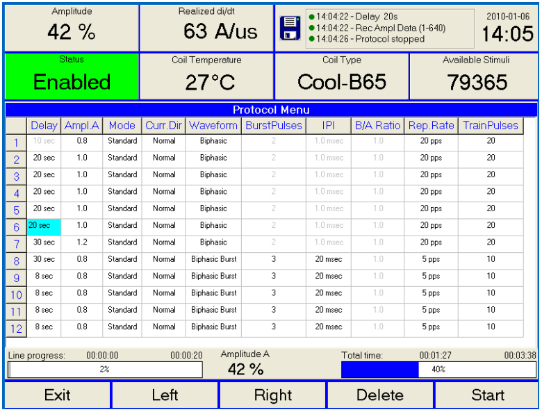
6. Using MEP Option motor evoked potential of TMS can be recorded
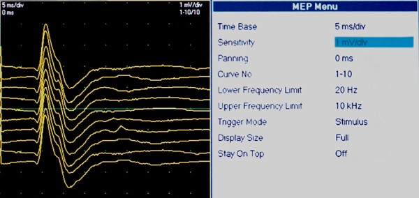
Figure 6: MEP signal recording using MEP option
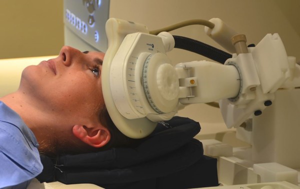
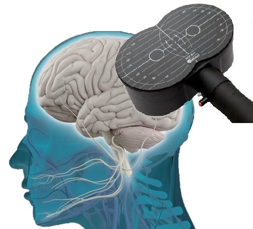
7. TMS-fMRI System Solution is a MR-compatible holder which can keep the coil in adjusted position.

Figure 7: Concurrent TMS/fMRI
TMS lab of NBML is one of the few centers in the world with the possibility of applying TMS during fMRI. All necessary facilities for this purpose (fMRI compatible coil, special room design next to imaging room and remotely controlling the TMS pulses, etc.) are provided in NBML.
Equipped with the frameless stereotaxic system, researches in TMS lab of NBML can use structural/functional brain images in defining TMS/tES stimulation targets individually with very high precision. This system enables researchers to obtain precise functional maps of different cortical regions for each subject.
Like any other type of study, placebo arm is crucial in TMS studies, specifically in clinical trials. Fortunately, TMS lab of NBML is equipped with specialized coils for both single and double blind studies.
Possible studies with TMS:
- Investigating Causal Brain-behavior Relationships and their Time Course with Very Good Spatial (few millimeters) and Temporal (a few tens of milliseconds) Resolution
- Navigated Magnetic and Electrical Stimulation of Cortex
- Possibility of single and double blind studies
- Concurrent TMS/fMRI
- Concurrent TMS/EEG
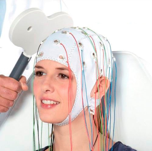
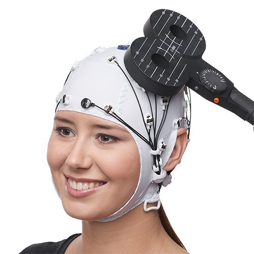
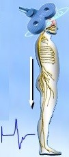
Figure 8: Concurrent EEG-TMS
List of Services (Top)
- MEP recording
- Implementation of the rTMS protocols for humans and rodents
- Determining the exact target points for magnetic stimulation using MRI images or using the MNI Atlas
- Recording the exact locations of stimulation, time and intensity of the applied pulse
- EEG recording during the sessions of magnetic stimulation
- Doing Functional MRI concurrent with TMS
- Running appropriate task with stimulation
- Motor and language mapping of the brain
Some Useful Links (Top)
- Professor Alvaro Pascual-Leone, Berenson-Allen Center for Noninvasive Brain Stimulation
- Professor Marom Bikson, University of New York
- Professor Mark S. George, Medical University of South Carolina
- Professor Michael Nitsche, Leibniz Research Centre for Working Environment and Human Factors
- Professor Vincent Walsh, University College London
- Professor Sven Bestmann, Institute of Neurology, University College London
- Combination of Stimulation and Monitoring
Experts in this field